Diagnosing CARP
BY CATALINA MATIZ, MD, AND ANDREA WALDMAN, MD
Frontline Medical News
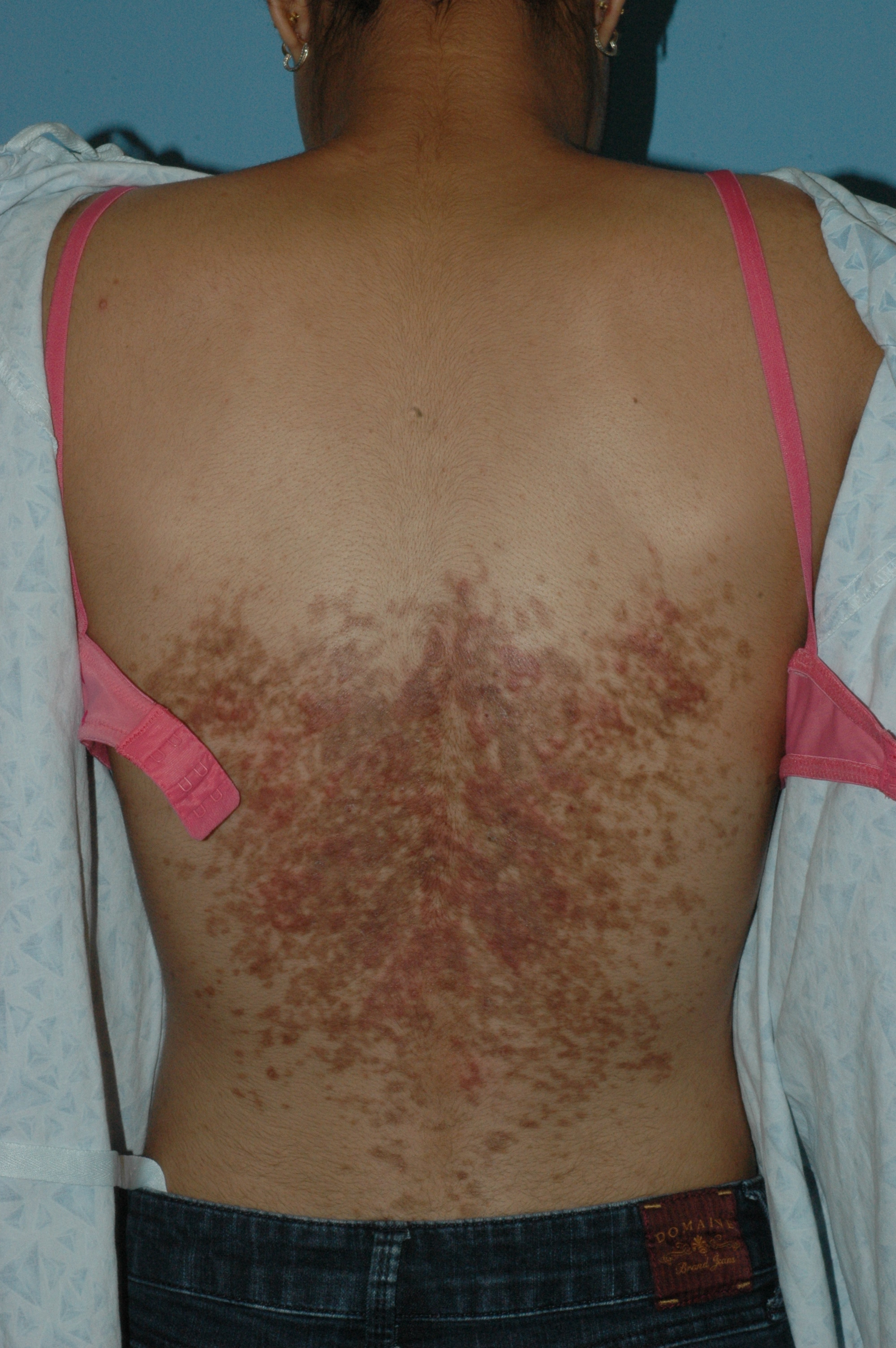
The distribution and morphology of our patient’s cutaneous eruption was highly suggestive of confluent and reticulated papillomatosis (CARP) of Gougerot and Carteaud, a rare dermatologic disorder characterized by epidermal changes (hyperkeratosis, scaling) and hyperpigmentation. The lesions classically begin as brown or red hyperpigmented hyperkeratotic or verrucous papules that spread centrifugally, coalescing into confluent plaques with reticulation at the periphery.1 CARP also may manifest as shiny papules or atrophic macules.1 Reports of scaly papules with rippling also are documented in the literature.2 Characteristically, the lesions frequently involve the inframammary region and the posterior trunk. Other less common sites of presentation include the abdomen, neck, axilla, face, and shoulders.1
Pathogenically, CARP results from disordered keratinization of the epidermis and increased melanosomes within the stratum corneum. The condition is chronic in nature and often intermittent, with a remitting-relapsing course. CARP is limited to cutaneous involvement, with no systemic manifestations. The lesions are typically asymptomatic; however, some individuals complain of mild pruritus.1 Primarily seen in adolescent patients, this dermatitis afflicts females twice as often as males, and occurs in all races.1
Several hypotheses concerning the etiology of CARP have been suggested in the literature; however, the definitive pathophysiology remains unknown. Suggested causes include defective keratinization, and infectious, endocrine, genetic, and environmental etiologies.3 In light of its clinical similarity to tinea versicolor and occasional clinical response to antifungal therapies, Malassezia has been postulated as a potential etiology for CARP. Potassium hydroxide examinations of the skin for fungal species are negative in most patients, and thus, this suggestion remains highly controversial.1 More convincing infectious culprits implicated include bacterial species such as Rhodococcus and Dietzia, papillomatosis previously isolated from skin scrapings of CARP patients. This is further supported by the successful treatment of CARP with antibiotic therapy.4 A highly controversial association between CARP and endocrine abnormalities such as insulin resistance and thyroid disease is well documented in the literature. Acanthosis nigricans and obesity may present concurrently with CARP as well; however, in most cases this association does not occur.3
The diagnosis of CARP is primarily clinical, formulated based on the presence of cutaneous lesions with the characteristic distribution and morphology. Biopsy and histopathology often are utilized to exclude other diagnoses. Davis et al. previously proposed the following five diagnostic criteria for CARP: involvement of the upper trunk and neck, clinical findings of scaly brown macules and patches (at least a portion of which appear reticulated and papillomatous), negative fungal staining of lesions, lack of response to antifungal therapy, and excellent response to minocycline therapy.5 These diagnostic criteria were validated by a retrospective analysis of CARP patients in Singapore.6
An extensive variety of cutaneous conditions bearing a resemblance to CARP were considered in the differential diagnosis, including acanthosis nigricans (AN), tinea versicolor, Darier disease, terra firma-forme dermatosis, prurigo pigmentosa, flagellate dermatosis, and dyskeratosis congenita.1 A common challenge for practitioners is distinguishing CARP from similar dermatoses, particularly AN. Differentiating AN from CARP cannot be accomplished based on the history of insulin resistance or increased body mass index alone, as these comorbidities may coexist with CARP.3 The clinical presence of reticulation peripherally, combined with a positive response to minocycline or azithromycin therapy, distinguish CARP from AN in most cases.
Other disorders commonly presenting in a similar distribution to CARP were further excluded based on the morphologic appearance of the lesions and further testing. Tinea versicolor was unlikely, because of the lack of response to antifungal agents and the absence of organisms on KOH examination. Furthermore, the morphologic characteristics of our patient’s lesions were inconsistent with the typical appearance of tinea versicolor – hypopigmented or hyperpigmented patches with overlying scale. Reticulated hyperpigmentation without papules or plaques may occur in dyskeratosis congenita.1 If the patient presents with reticulated hyperpigmentation in addition to pruritus, prurigo pigmentosa may be considered in the differential.4 The presentation of slightly verrucous “dirtlike” plaques refractory to cleansing with soap and water alone, should prompt the consideration of terra firma-forme dermatosis. Terra firma-forme dermatosis, a benign disorder of keratinization, may be confirmed at the bedside upon the removal of the plaques with a 70% alcohol swab.7 Flagellate erythema, characterized by the presence of linear streaks of erythema or brown pigmentation, often presents in a similar distribution to CARP. This condition occurs mostly in individuals treated with bleomycin or other chemotherapeutic agents, and spontaneously resolves.1 Darier disease classically presents as hyperkeratotic papules coalescing into plaques in seborrheic areas, and may manifest in the same distribution as CARP. Palmoplantar pits and nail changes (blue-red striations and distal V-shaped nicking) may be present in the former.1
A variety of treatments for CARP are documented in the literature, including antibiotic therapy with minocycline or azithromycin, topical salicylic acid, urea or lactic acid–containing emollients, and oral or topical retinoids.1,3 A substantial proportion of patients respond to oral minocycline (100 mg twice daily). At the time of initial evaluation, our patient was prescribed a 6-week course of systemic minocycline (100 mg twice daily), with subsequent improvement.
References
1. Am J Clin Dermatol. 2006;7(5):305-13.
2. Postepy Dermatol Alergol. 2014 Oct;31(5):335-7.
4. Br J Dermatol. 2006 Feb;154(2):287-93.
5. Ann Dermatol. 2014 Jun;26(3):409-10.
6. Am J Clin Dermatol. 2015 Apr;16(2):131-6.
7. Clinical Experiment Dermatol. 2012;37(4):446-7.
Dr. Matiz is assistant professor of dermatology at Rady Children’s Hospital San Diego, University of California, San Diego. Dr. Waldman is a clinical research fellow at the hospital. Dr. Matiz and Dr. Waldman said they had no relevant financial disclosures.


