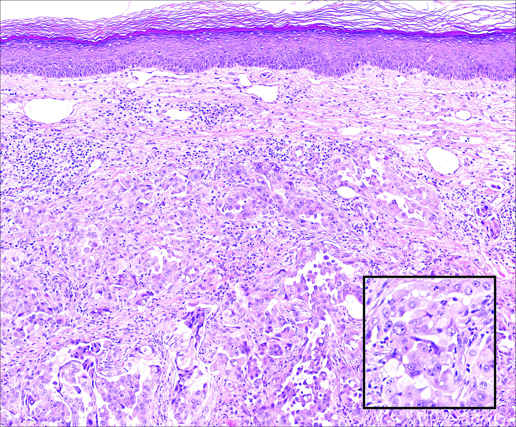
Primary sebaceous carcinoma also can mimic metastatic adenocarcinoma within the skin and is histologically similar to metastatic adenocarcinomas. The most distinguishing feature is sebaceous differentiation characterized by sebocytes, which have a vacuolated cytoplasm giving the nucleus a scalloped appearance, frequently with adjacent ductlike structures (Figure 5). Epidermotropism sometimes is present in sebaceous carcinomas but cannot be relied on as a distinguishing feature. Immunohistochemical analysis also is a helpful tool; these tumors typically are positive for p63 and podoplanin, distinguishing them from negative-staining metastatic adenocarcinomas.10,11
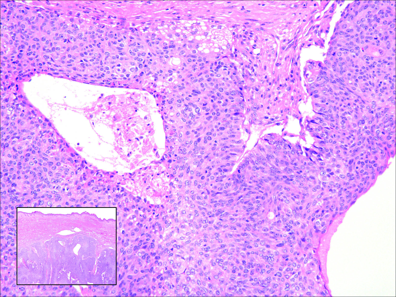
Figure 5. Within the dermis is a dense collection of atypical cells (inset [H&E, original magnification ×20]) with an unaffected overlying epidermis. At higher magnification, the atypical cells are elongated with abundant eosinophilic cytoplasm. Ductlike structures and vacuolated cytoplasm are characteristic of sebaceous carcinoma (H&E, original magnification ×400).