Take-Home Points
- TSTC-PLFD is a rare hyperextension wrist injury characterized by fracture of both the scaphoid and the capitate and rotation of the proximal bone fragment of the capitate.
- TSTC-PLFD is associated by a complex ligamentous injury of the wrist.
- Impaction of the wrist in extension seems to be the most important predictor of this injury.
- Optimal treatment for TSTC-PLFD is open reduction, anatomical alignment, and ligamentous and osseous stabilization.
- The most important complications of scaphoid and capitate fractures and PLFD are osteonecrosis and nonunion.
Trans-scaphoid transcapitate (TSTC) perilunate fracture-dislocation (PLFD) is a rare hyperextension wrist injury characterized by fracture of both the scaphoid and the capitate and rotation of the proximal bone fragment of the capitate.1 Isolated capitate fractures with or without rotation of its proximal fragment have been well described.2,3 Obviously, this specific type of injury represents just the osseous part of a more complex ligamentous wrist injury.2,3
TSTC-PLFD was first described by Nicholson4 in 1940. In 1956, Fenton5 coined the term scaphocapitate syndrome, which became widely known. With PLFD, accurate diagnosis may be delayed. Usually, only the scaphoid fracture is identified by radiologic examination, and thus the severity of the injury is underestimated and appropriate treatment delayed.3,6,7 The English literature includes only case reports and small series on this rare perilunate injury.6-9 In this article, we report the case of an adult with TSTC-PLFD. We describe the radiographic and intraoperative findings, review the current surgical principles for reduction and stabilization of this injury, and assess the clinical and radiologic outcomes. The patient provided written informed consent for print and electronic publication of this case report.
Case Report
A 32-year-old man sustained an isolated injury of his right (dominant) hand after falling from a height of 6 feet and landing on his outstretched right arm with the wrist in extension.
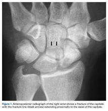
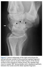
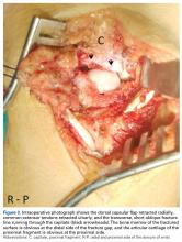
Physical examination at admission revealed swelling over the dorsum of the wrist and pain on palpation. Radiographs showed a fracture of the waist of the scaphoid (Figure 1). In addition, the capitate was fractured with the proximal fragment rotated 180° (Figure 1, Figure 2). A small avulsion fracture on the dorsal surface of the wrist was obvious as well (Figure 2). A perilunate injury was diagnosed and surgical treatment recommended.With the patient under general anesthesia and a humerus tourniquet applied, an external fixator was placed for spanning of the wrist joint. The dorsal aspect of the wrist joint was approached through a midline longitudinal 5-cm incision, centered over the Lister tubercle. For adequate exposure of the dorsal wrist, a flap of the dorsal capsule was raised with the apex at the triquetrum and a radial broad base, as previously described.9 An avulsion fracture at the insertion of the dorsal capsule to the triquetrum was observed. The dorsal surface of the hamate and lunate showed a small area of bone contusion with hemorrhagic infiltration. The scapholunate and lunotriquetral ligaments were intact. The proximal fragment of the capitate was identified deep into the space between the lunate and distal capitate fragment; the articular surface of the bone fragment was rotated 180° distally (Figure 3).
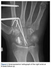
Distraction was applied through the external fixator, and the bone fragment was removed from the surgical site. The cartilaginous surface was scratched, but no chondral flap or defect was observed. Hematoma and debris were removed, and the bone fragment was restored to its anatomical position. Two 1.6-mm Kirschner wires (K-wires) were inserted in a distal-to-proximal direction to stabilize the capitate fracture without engaging the lunate. The scaphoid fracture was reduced and stabilized with an antegrade double-threaded compression screw. Then, both K-wires were advanced proximally, engaging the lunate, to try to enhance midcarpus anteroposterior stability (Figure 4). The scapholunate and lunotriquetral intervals were stable. Last, the wound was sutured in layers, and the external fixator was locked with the wrist in 0° of flexion-extension and 0° of radioulnar deviation.Skin sutures were removed 2 weeks after surgery, K-wires 6 weeks after surgery, and the external fixator 8 weeks after surgery. At 8 weeks, radiographs showed healing of both fractures, scaphoid and capitate. The patient was allowed gradual passive and active-assisted range-of-motion exercises of the wrist at 8 weeks, and he returned to work 3 months after surgery. At 12-month follow-up, all fractures were completely healed, and the wrist was stable and pain-free.
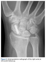
At 24-month follow-up, the patient was asymptomatic, had no ulnar translation of the right wrist joint, and showed full range of pronation-supination, a 10° lag of wrist flexion, and a 20° lag of extension in comparison with the left wrist. Mayo wrist score was excellent (95 points). Radiographs of the right wrist showed fracture healing and ligamentous stability of the carpal joints (Figure 5).
